CBCT Scan in Chișinău
A CBCT scan is a three-dimensional X-ray that shows the jaws, teeth, roots and surrounding tissue. On a standard X-ray everything overlaps in one plane, while a 3D dental scan lets the doctor look at every root and every area of bone from all sides. We order it before implant placement, for complex root canal treatment, before wisdom tooth removal and whenever the doctor needs an exact picture.
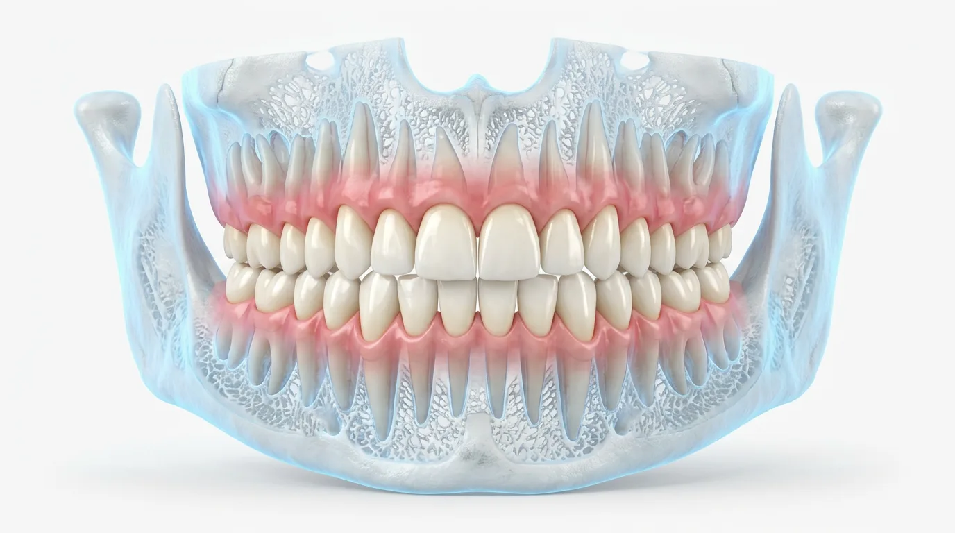
What is a CBCT scan of the teeth
Dentistry uses cone beam computed tomography (CBCT). The scanner makes one turn around the patient's head and captures a series of images in a few seconds, and software builds a three-dimensional model from them. On screen the doctor can slice the model in any plane, measure the height and width of the bone to a fraction of a millimetre, and trace the nerve in the lower jaw and the borders of the maxillary sinus. In our practice the main advantage over two-dimensional radiography is that we see slices without overlapping structures.
A CBCT scan does not replace an examination, it supplements it. We order a scan when the choice of treatment or the safety of an operation depends on knowing the anatomy exactly. For everyday caries diagnosis a periapical (close-up) or panoramic X-ray is usually enough, and we take those as part of dental X-rays. We follow a general principle: use the method with the lowest radiation dose that answers the clinical question.

When you need a dental scan
Most often a 3D dental scan is ordered before dental implant placement. From the scan the surgeon assesses the volume of bone, chooses the implant size and angle, and sees the distance to the nerve and the sinus. We plan every implant from a CBCT scan, especially when there is little bone or additional operations are needed. If bone is lacking, the scan shows whether a sinus lift or ridge augmentation (rebuilding the bone) is required.
The second common indication is endodontics (root canal treatment). With complex root anatomy, a suspected extra canal, crack or perforation (a hole in the root wall), or inflammation at the root tip that does not show on a standard X-ray, the scan helps to make an exact diagnosis. It is needed before root canal retreatment, when the doctor has to understand why the first treatment failed. A jaw scan is also done before complex removal of impacted teeth (stuck in the bone), when a cyst is suspected, after injuries and to assess the temporomandibular joint (the jaw joint).
CBCT scan vs standard X-ray: what is the difference
Parameter | CBCT scan (3D) | Panoramic and periapical (close-up) X-ray |
|---|---|---|
Image | three-dimensional, slices in any plane | flat, structures overlap |
What it shows | bone, roots, canals, nerve and sinus in 3D | general view of the jaws or 1-3 teeth |
Measurements | precise, in millimetres | approximate |
Radiation dose | higher, depends on the scanned area | lower |
When it is ordered | implants, complex endodontics, surgery | caries, treatment control, first examination |
Our price | 700 MDL | 200 / 100 MDL |
A standard X-ray answers the question of whether there is a problem. A scan shows where exactly it is and how large it is. That is why we start with the simpler examination and order a CBCT scan only when treatment cannot be planned without it. The radiation dose of dental tomography is higher than that of a panoramic X-ray, which is why we order it only when it is needed.
When you do not need a scan
If the doctor only needs to see caries, the quality of an old filling or the result of a root canal treatment, a periapical X-ray is enough. For a general overview of all the teeth, an assessment of wisdom teeth and the state of the bone in gum disease, a panoramic X-ray is suitable. We do not scan just in case and do not repeat a scan without a medical reason.
What the scan gives you
The size and position of the implant are chosen from the real volume of bone, not by guesswork.
The surgeon sees the nerve, the sinus and the roots of neighbouring teeth in advance.
The scan shows extra canals, cracks and areas of inflammation.
A repeat scan shows how an implant has integrated or how a lesion at the root has healed.
Cost of a jaw CBCT scan
Our CBCT scan costs 700 MDL. If you are treated with us, a follow-up scan, for example after implant placement or root canal treatment, is free. A consultation with a specialist doctor is also free: the doctor goes through the scan with you and explains what treatment you need and how much it will cost. For comparison, a panoramic X-ray costs 200 MDL and a periapical X-ray costs 100 MDL. The doctor sets out the final plan and estimate before treatment begins.
How the scan is done
At the consultation the doctor decides which area to scan: one jaw, a section of several teeth or both jaws.
You remove glasses, earrings, removable dentures and other metal objects around the head and neck.
You stand or sit, rest your chin on the support and lightly bite on a special holder.
The machine makes a turn around your head, and you need to stay still for a few seconds.
The software builds a three-dimensional model, which the doctor studies in different planes.
The doctor shows you the scan, explains the findings and proposes a treatment plan.
How long the scan takes
The scan itself takes a few seconds, and the whole procedure with preparation takes about 10-15 minutes. No preparation is needed in advance, and you can eat and drink as usual. The doctor studies the result on the same day. In complex cases, for example before All-on-4 implants, extra time may be needed for planning. The scan file is saved, so it can be used at the next stages of treatment and compared with follow-up scans.
What happens if treatment starts without proper diagnostics
Without data on the volume of bone, an implant can turn out too long or too wide.
When working on the lower jaw it is vital to know exactly where the nerve runs.
An untreated extra canal is a common cause of repeated inflammation.
An inaccurate diagnosis leads to redoing the work, extra visits and extra costs.
Who needs a scan most
A scan matters most for those planning implants after teeth were lost long ago, for those facing removal of a complex wisdom tooth close to the nerve, for those whose tooth has hurt for a long time after root canal treatment, and for those with a shadow at the root on a standard X-ray. In such cases a CBCT scan helps to avoid unnecessary procedures and to choose a gentler option. Before wisdom tooth removal, for example, the surgeon uses the scan to see how close the roots are to the nerve and plans the removal so as to lower the risk of complications. In root canal treatment the endodontist uses the scan to determine the number and shape of the canals and to choose the instruments.
Before the examination, bring your old X-rays and medical records if you have them: the doctor will compare them with the new scan and assess how the condition of your teeth and bone has changed. If you have already had a scan elsewhere, bring the file on a disc or a flash drive. The doctor will tell you at the consultation whether it is suitable for planning or whether a new scan is needed.
Frequently asked questions about CBCT scans
Don't put off your dental health


