Dental X-Ray in Chișinău
A dental X-ray shows what a visual check cannot: decay between teeth and under fillings, the condition of roots and canals, inflammation at the root tip, the position of wisdom teeth and the level of bone around the teeth. We take two types of images: a panoramic X-ray (OPG) and a close-up (periapical) image on a digital sensor. Our doctor chooses the type for your particular question and goes through the result with you at the appointment.
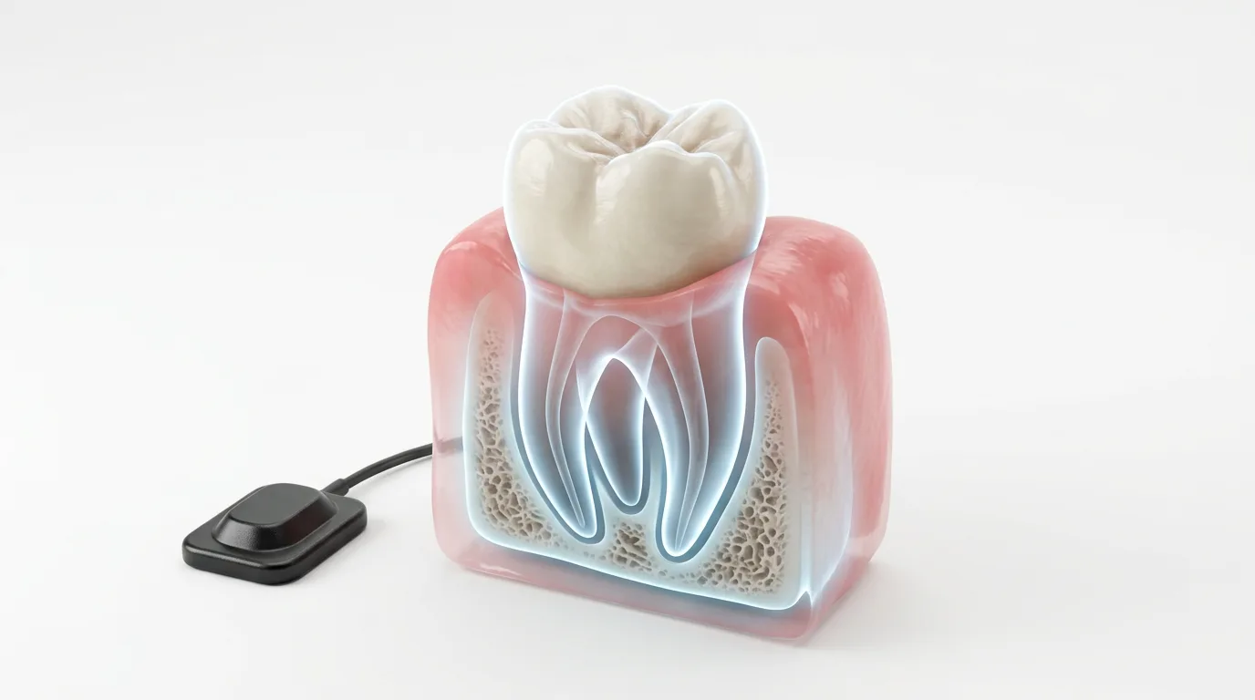
Types of dental X-rays
A panoramic X-ray is an overview of both jaws in one picture. It shows all the teeth, including unerupted ones, the roots, the maxillary sinuses, the jaw joints and the general bone level. The procedure is called orthopantomography, and the image is an orthopantomogram (OPG). We take it at the first examination, before planning complex treatment, for gum disease and to assess wisdom teeth.
A close-up (periapical) image shows one to three teeth in detail. We take it with a digital sensor: the sensor is placed in the mouth next to the tooth, and the image appears on the computer screen at once. On screen we can enlarge it, adjust the contrast and see details that a panoramic image does not show. Close-up images are needed during root canal treatment, to check fillings and when we suspect decay on the contact surfaces between teeth.
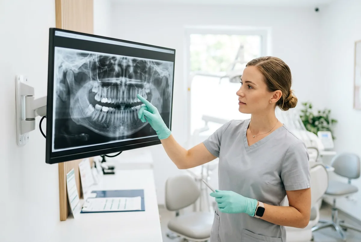
When you need a dental X-ray
We order an image when an examination alone cannot tell us what is happening with a tooth. Typical situations: a tooth hurts and there is no visible cause; we suspect decay between teeth or under an old filling; we need to assess a root before root canal treatment and check the quality of the filling afterwards; your gums bleed and we want to check the bone around the teeth; an extraction, an implant or a prosthesis is planned.
For gum disease, the panoramic image shows the bone level around all the teeth, and close-up images clarify the condition of individual areas. In gum treatment (periodontology) we use these data to plan treatment and then to compare the result. Before removing a difficult wisdom tooth we usually start with a panoramic image, and if the roots lie close to the nerve, we order a CBCT scan (3D dental scan).
Panoramic and close-up dental X-ray: what is the difference
Parameter | Panoramic X-ray | Close-up X-ray |
|---|---|---|
What it shows | both jaws, all teeth, sinuses, joints | one to three teeth in detail |
Detail | overview | high, small root details are visible |
When we order it | first examination, treatment plan, wisdom teeth, gums | root canal treatment, decay, checking fillings |
How it is taken | the machine rotates around the head | the sensor sits inside the mouth |
The two images complement each other. The panoramic image gives the overall picture, and the close-up image answers a specific question about one tooth. To assess inflammation at the root tip, for example, a close-up image is usually more informative, because the picture is larger and less distorted. If a two-dimensional image is not enough, for example with complex root anatomy or a lesion that persists after treatment, we may order a CT scan.
When you do not need an X-ray
We do not take an X-ray without a medical reason. If all you need is plaque removal or polishing of a filling, no image is required. How often preventive images are taken to look for decay depends on individual risk: people with multiple cavities have them more often, and people at low risk less often. We take an X-ray only when the image will change the treatment. Tell us if you are or may be pregnant. In that case we take an image only when it is necessary.
What a dental X-ray gives you
We see decay between teeth, under fillings and under crowns.
We check the length and density of the filling in every canal.
The image shows the bone level in gum disease and before implantation.
The panoramic image is the basis for the overall plan and the order of visits.
Dental X-ray price in Chișinău
An overview of both jaws and all teeth.
A digital image of one to three teeth in detail.
The doctor goes through the image and explains the treatment options.
The specialist consultation is free: the doctor shows the image on screen and explains what it shows. If treatment needs a three-dimensional picture, we carry out a CT scan for 700 MDL, and the follow-up CT is free for our patients. Every image is part of the overall treatment plan, and its cost is fixed in advance.
How a dental X-ray is taken
The doctor decides which image is needed: panoramic or close-up.
You take off earrings, glasses, removable dentures and any jewellery around the head and neck.
If needed, we use a protective apron or collar.
You stand at the machine, bite on the holder and stay still for a few seconds while the machine moves around your head.
The sensor is placed in the mouth next to the tooth, and the image takes a fraction of a second.
The image appears on the screen at once, and the doctor explains what it shows.
How long a dental X-ray takes
A close-up image takes a couple of minutes right in the dental chair, so we often take it during treatment, for example to measure the length of the canals. The panoramic image itself takes a few seconds, and the whole procedure, with preparation, takes about five minutes. No special preparation is needed, and you can eat and drink as usual. The result is ready at once. It can be saved and used for comparison at later visits.
What happens if treatment starts without an X-ray
Decay between teeth and under fillings stays invisible for a long time and destroys the tooth from inside.
An area of inflammation at the root tip can develop for years without pain.
Without a check on the image it is hard to be sure the canals are filled to the end.
Without a panoramic image, problems of neighbouring teeth and bone can be missed.
Is a dental X-ray safe
Modern digital images give a low radiation dose, especially close-up images on a sensor. Even so, we order any X-ray only when it is indicated and keep the number of images to a minimum. We keep the dose low: we use digital sensors, take only the images we need and use a protective apron or collar when needed. With children and teenagers we are stricter still: images only for clear indications and always with protective equipment. If you have had images taken recently at another clinic, bring them: we will assess whether a repeat examination can be avoided.
Regular images matter most for people with decay under old fillings, crowns or implants. Comparing images over several years shows whether the bone is stable and whether new lesions have appeared. This lets us spot a problem in time and treat it before pain begins. Before a wisdom tooth removal, the panoramic image shows the position of the roots and the neighbouring teeth, and when in doubt we add a CT scan.
Dental X-ray FAQ
Don't put off your dental health


