updated
November 12, 2026
...
read
Periapical Granuloma: Causes, Symptoms and Treatment of a Tooth Granuloma
A periapical granuloma, or tooth granuloma, is a small focus of inflamed tissue at the tip of the root. It forms in response to infection inside the tooth. Most often the granuloma does not hurt and is visible only on an X-ray. We treat it first of all through the tooth: we clean and fill the root canals, after which the small focus often heals.

What a periapical granuloma is
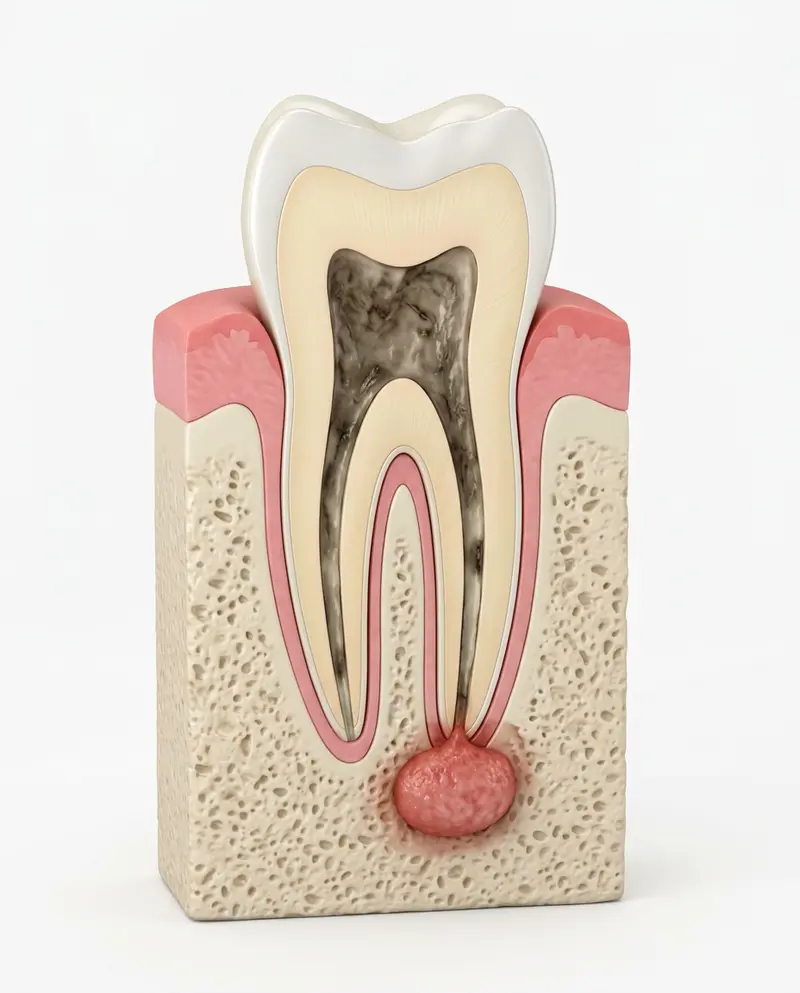
A periapical granuloma is a defensive reaction of the body. It is a focus at the tip of the root. When the nerve dies because of deep decay or an injury, bacteria from the canal reach the tip of the root. The body walls them off with dense granulation tissue, that is, young inflamed tissue. A small focus appears in the bone.
Unlike a cyst, a dental granuloma has no wall of its own made of epithelium, the lining of cells on the inside. We regard it as a stage from which a cyst can develop over time.
A tooth granuloma is, in essence, a signal. There is an infection inside the tooth that the body has not fully dealt with.
Causes
- deep decay that has reached the nerve;
- an injury to the tooth after which the nerve died;
- root canals that were poorly treated or left untreated;
- a crack in the root.
The early signs of decay, before it reaches the nerve, are described in the article on signs of cavities you should not ignore.
Dental granuloma symptoms
Often there are none. In our practice, the focus is often a chance finding: a person comes for a routine check-up or before prosthetics, the tooth causes no trouble, and the X-ray shows a dark spot at the tip of the root.
What the patient may feel
- slight pain or discomfort when biting
- darkening of the tooth after an injury or old treatment
- a small pimple on the gum, sometimes with pus
- a feeling that the tooth has become taller than the others
What we see on the X-ray
- a dark spot at the tip of the root
- the condition of the filling in the canals, if the tooth has been treated
- how far the focus has destroyed the bone
- changes compared with older images
In a flare-up there is severe pain, swelling and a raised temperature.
Warning signs
- swelling of the gum or cheek that is increasing;
- a raised temperature and weakness;
- difficulty opening the mouth or swallowing.
In these cases you need a doctor on the same day. This text is for general information only and does not replace a dentist's examination.
Cyst and granuloma of the tooth: how to tell them apart
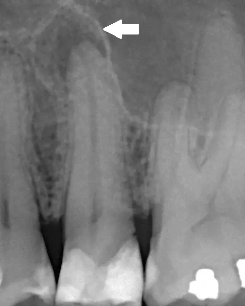
On an X-ray both a granuloma and a cyst look like a dark spot at the tip of the root. An ordinary image cannot always tell them apart reliably. To assess the size and borders of the focus, we can order a CBCT scan (cone-beam computed tomography, a 3D scan). But even that gives only a preliminary assessment. According to international studies, the type of tissue is finally confirmed by examination under a microscope when the focus is removed.
This matters for one practical reason. Whether root canal treatment is enough or surgery is needed depends on the size and character of the focus.
Found a focus at the root on an X-ray?
We will explain what the image shows and how to save the tooth.
Tooth granuloma treatment
Root canal treatment
We remove the dead nerve and the bacteria, rinse the canals and seal them tightly with a filling.
Retreatment
If the canals have already been filled but the infection remains, we carry out root canal retreatment, with magnification where necessary.
Follow-up images
After a few months we check whether the focus is shrinking.
Apicoectomy
If the focus does not go away after treatment, or the canals cannot be fully cleaned, we surgically remove the tip of the root together with the focus.
Tooth extraction
We remove the tooth only when it cannot be saved.
What to expect from root canal treatment
Treatment is done under local anaesthesia. We isolate the tooth, open access to the canals, clean them with instruments and solutions and fill them tightly. Sometimes two visits are needed, especially if there is a lot of infection in the canal or old filling material. In the first days the tooth may ache slightly when you bite. This is a normal reaction.
We judge the result not by how you feel but by the images: the bone around the root recovers over months. According to international studies, the outcome of retreatment is influenced by whether there was a focus before treatment, by how precisely the filling reaches the tip of the root, and by the quality of the crown restoration. That is why after treatment we immediately plan how to restore and protect the tooth itself.
Granuloma after tooth extraction
Sometimes after a tooth is removed, inflamed tissue is left in the bone. That is why, during an extraction, we clean the socket thoroughly. If the socket hurts for a long time, bleeds or discharges pus, come to see us: a focus can keep the inflammation going.
Treatment prices at Cristi Dent
Prices are valid as of 19 October 2026. We determine the scope of treatment after the examination and the X-ray. More about the price is on the root canal retreatment page.
Bothered by a tooth that was treated some time ago?
Book a free consultation: we will check the canals and the bone around the root.
Where to go
Has an X-ray shown a focus at the root? Or does a tooth that was treated some time ago bother you? Start with a consultation. Sometimes root canal treatment is not enough. Then we carry out an apicoectomy to save the tooth. We do it at Cristi Dent in Chișinău.
Sources
- StatPearls. Odontogenic Cysts www.ncbi.nlm.nih.gov/books/NBK574529
- StatPearls. Oral Facial Infection of Dental Origin: A Guide for the Medical Practitioner www.ncbi.nlm.nih.gov/books/NBK542165
- Pitcher B et al. Binary Decision Trees for Preoperative Periapical Cyst Screening Using Cone-beam Computed Tomography. J Endod. 2017;43(3):383-388 pubmed.ncbi.nlm.nih.gov/28231977
- Ng YL et al. Outcome of secondary root canal treatment: a systematic review of the literature. Int Endod J. 2008;41(12):1026-1046 pubmed.ncbi.nlm.nih.gov/19133093
Images in the article: a focus at the root on an X-ray: Michele Gardini, CC BY-SA 3.0, Wikimedia Commons.
Consequences of a tooth granuloma without treatment
A granuloma will not go away on its own while there is infection in the tooth. Without treatment, the bone around the root may gradually be destroyed, the granuloma may turn into a cyst, a flare-up with purulent inflammation may occur and the tooth may be lost.
After treatment
- come for follow-up images at the times we set;
- restore the tooth with a filling or a crown, as we advise;
- do not chew hard food on this tooth until it has been restored.
Frequently asked questions
It keeps the infection going in the bone, can flare up with swelling and pus, turn into a cyst and lead to the loss of the tooth.
You can live with it for years without symptoms, but the infection does not go anywhere. It is better to treat it before it flares up.
A cyst has a wall and a cavity of its own, while a granuloma does not. On an ordinary X-ray they look similar, and at the start the treatment is the same: through the root canals of the tooth.
Usually one or two visits for root canal treatment. Then, after a few months, we take a follow-up image. If an apicoectomy is needed, it is a separate visit.
A consultation with a specialist doctor at Cristi Dent is free. We will study the image and tell you whether root canal treatment is enough.
ARTICLE AUTHOR
Carries out root canal treatment and retreatment, restoration of badly damaged teeth and preparation of teeth for further prosthetics.