updated
November 12, 2026
...
read
Dental Cyst: Symptoms and Whether the Tooth Can Be Saved

A dental cyst is a cavity with fluid contents and a wall that forms in the bone at the tip of the root. Most often it appears after the nerve has died. It does not hurt for a long time, and we usually find it by chance on an X-ray. In many cases the tooth can be saved: we treat the root canals and, if necessary, remove the cyst surgically.

Dental cyst: symptoms and whether the tooth can be saved
What a cyst at the tip of the root is, why it does not hurt for a long time, how it is found on an X-ray and how it is treated: root canals, a follow-up image, removal of the root tip (apicoectomy). Cristi Dent, Chișinău.
What a dental cyst is and why it is dangerous

A dental cyst often does not hurt for years, and in many cases the tooth can be saved. In short, a dental cyst is the result of chronic infection inside the tooth. The nerve dies because of deep decay or an injury. Bacteria then pass through the canal to the tip of the root. Young inflamed tissue grows around the focus, and this is how a granuloma forms. In some cases a cavity with an epithelial wall, that is, a thin lining of cells, forms inside it. This is the cyst.
According to international studies, this kind of cyst is more common than all other cysts of dental origin. But far from every dark spot at the root turns out to be a cyst. And even a large focus does not always mean the loss of the tooth.
We take the second figure from international studies of lesions at the root, and the third from a clinical series in which patients were followed for 2 to 10 years.
A cyst at the root is called a radicular cyst or a periapical cyst. In our practice, it is more common in the upper jaw. Is a tooth cyst dangerous? It is dangerous not because it grows fast but because it grows unnoticed:
- it gradually destroys the bone around the root;
- it can fill with pus and turn into an abscess with swelling of the face;
- in teeth of the upper jaw it sometimes affects the maxillary sinus, the cavity in the bone above the teeth;
- a large cyst weakens the jaw and makes future dental implants harder.
Why a radicular cyst appears
The main cause is a dead nerve, with bacteria left in the canals afterwards. This happens:
- with deep decay that was not treated;
- after an injury to the tooth, even without a visible chip;
- after root canal treatment, if the infection in the canals remained.
Does a dental cyst hurt
Often it does not. While the focus is quiet, you feel nothing. We find the cyst on an X-ray during a routine check-up, when preparing for prosthetics, or on an image taken for a completely different reason. These may put you on your guard:
- slight discomfort when biting
- a feeling of pressure around the tooth
- a pimple on the gum, from which pus sometimes comes out
- darkening of a tooth that was once treated or injured
In a flare-up there is pain, swelling and a raised temperature. This is already a reason to see a dentist the same day.
Warning signs
- swelling of the cheek or under the jaw that is increasing;
- a raised temperature and general malaise;
- difficulty opening the mouth or swallowing;
- swelling near the eye.
The material in this article is for general information and does not replace an examination by a doctor.
How the diagnosis is made
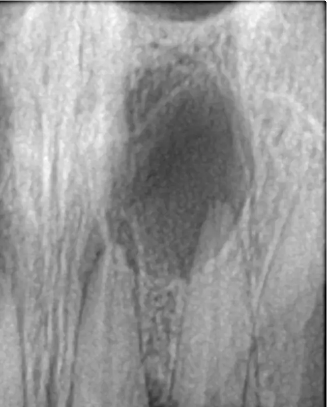
On an X-ray a cyst looks like a rounded dark spot with clear edges at the tip of the root. An ordinary image does not always tell a cyst from a granuloma. So where necessary we take a CBCT scan (3D dental scan). The exact type of formation is confirmed by examining the tissue under a microscope after removal.
It depends on the size and character of the focus whether root canal treatment is enough or surgery is needed. That is why we compare images before and after treatment and do not rely only on how you feel.
Granuloma
- dense inflamed tissue at the tip of the root
- no wall of its own
- often heals after good-quality root canal treatment
Cyst
- a cavity with fluid and a wall of epithelium
- can grow for years
- if the wall is dense, it may remain after root canal treatment, and then we remove it
Found a dark spot at the root on an X-ray?
We will explain what the focus is and suggest how to save the tooth.
How a dental cyst is treated
We choose the approach by the size of the formation, the condition of the tooth and whether it has been treated before. Below is the typical order.
Root canal treatment or retreatment
We clean and fill the canals, removing the source of infection.
Follow-up image
After a few months we check whether the focus is shrinking. Bone recovers slowly.
Apicoectomy
If the focus does not shrink, or the canals cannot be treated, we remove the cyst together with the tip of the root.
Tooth extraction
We carry out tooth extraction and clean out the cyst if the tooth is destroyed and cannot be saved.
Root canal treatment is part of our endodontics service, and the surgical stages are handled by our surgeon.
At consultations we are often asked whether a tooth has to be removed if a cyst has been found on an X-ray. In our practice, there is no need to rush. First we look at whether the tooth was treated before, whether the canals can be accessed and how much bone is left around the root. If the tooth can be restored, we start with root canal treatment and compare follow-up images. We leave extraction for when the tooth really cannot be saved.
What to do to make a dental cyst go away
Folk rinses and tablets will not dissolve a cyst, however convincing the advice on forums sounds. Only one thing gives a chance of shrinking: removing the infection inside the tooth with good-quality root canal treatment. Small foci often heal after this, and we see it on follow-up images. A cyst with a dense wall may not disappear. Then we remove it.
Can a cyst in a tooth go away on its own
No. Without treatment, the source of infection inside the tooth does not go anywhere. The focus slowly grows and destroys the bone around the root. The absence of pain only postpones the moment when you find out about the focus.
Will antibiotics help
Not on their own. An antibiotic can relieve a flare-up for a while, but it does not get inside the dead tooth where the bacteria live. When the course ends, the focus stays where it was. According to international studies, the source of an acute infection at the root is removed first. Antibiotics are indicated when the infection spreads or there are general symptoms. That is why we prescribe tablets only in such cases and together with root canal treatment, not instead of it.
Can a dental cyst come back
Yes. The focus can form again in the same place. This happens if the infection in the canals remained or the cyst was not removed completely. A so-called residual cyst appears if part of it was left in the bone after the tooth was removed. That is why after treatment we always look at follow-up images.
Treatment prices at Cristi Dent
Prices are valid as of 19 October 2026. X-rays and root canal retreatment before an apicoectomy are paid separately. More details on the apicoectomy page.
Want to save a tooth with a cyst?
Book a free consultation: we will tell you whether root canal treatment is enough.
Where to go
An X-ray may have shown a dark spot at the root. Or a pimple on the gum may bother you. Either way, start with a consultation at Cristi Dent in Chișinău. We save the tooth whenever possible. If the focus does not shrink after root canal treatment, we carry out an apicoectomy with removal of the cyst.
Sources
- StatPearls. Odontogenic Cysts www.ncbi.nlm.nih.gov/books/NBK574529
- StatPearls. Oral Facial Infection of Dental Origin: A Guide for the Medical Practitioner www.ncbi.nlm.nih.gov/books/NBK542165
- NHS. Dental abscess www.nhs.uk/conditions/dental-abscess
- Johnson NR et al. Frequency of odontogenic cysts and tumors: a systematic review. J Investig Clin Dent. 2014;5(1):9-14 pubmed.ncbi.nlm.nih.gov/23766099
- Fernandes M, de Ataide I. Nonsurgical management of periapical lesions. J Conserv Dent. 2010;13(4):240-245 pubmed.ncbi.nlm.nih.gov/21217952
- Caliskan MK. Prognosis of large cyst-like periapical lesions following nonsurgical root canal treatment. Int Endod J. 2004;37(6):408-416 pubmed.ncbi.nlm.nih.gov/15186249
- Cope AL et al. Systemic antibiotics for symptomatic apical periodontitis and acute apical abscess in adults. Cochrane Database Syst Rev. 2024;5:CD010136 pubmed.ncbi.nlm.nih.gov/38712714
Images in the article: a focus at the root on an X-ray: Shaimaa Abdellatif, CC BY-SA 4.0, Wikimedia Commons.
Consequences of a dental cyst without treatment
If you leave everything as it is, the consequences build up over the years. The destruction of bone goes further, the risk of acute inflammation grows and the neighbouring teeth suffer. It is harder to restore this area later, and sometimes by then the tooth can no longer be saved.
After treatment
- come for follow-up images at the times we set;
- do not skip an image even if nothing hurts;
- restore the tooth with a filling or a crown, as we advise.
Frequently asked questions
If neither the tooth nor the cyst is treated, the focus keeps growing slowly, destroys the bone and can fill with pus. But removal is not the only way out: we often save a tooth with a cyst by treating its canals.
In many cases, yes. First we treat or retreat the canals, and if the focus does not shrink, we carry out an apicoectomy.
By follow-up images. The bone around the root recovers over months, so we compare new images with the first ones. The absence of pain is not an indicator in itself.
We carry out the apicoectomy under local anaesthesia, so there is no pain during the operation. For a few days afterwards there may be swelling and moderate discomfort, and we will tell you how to relieve them.
A consultation with a specialist doctor at Cristi Dent is free. We will study the image, assess the focus and suggest a treatment plan.
ARTICLE AUTHOR
Carries out root canal treatment and retreatment, restoration of badly damaged teeth and preparation of teeth for further prosthetics.